- Anterior Talo-Fibular Ligament (ATFL): running anteriorly from the lateral malleolus to the talusPosterior Talo
- Fibular Ligament (PTFL): running posteriorly from the lateral malleolus to the posterior aspect of the talus
- Calcaneo-Fibular ligament (CFL): running from the lateral malleolus to the lateral aspect of the calcaneous, in the middle between the ATFL and the PTFL
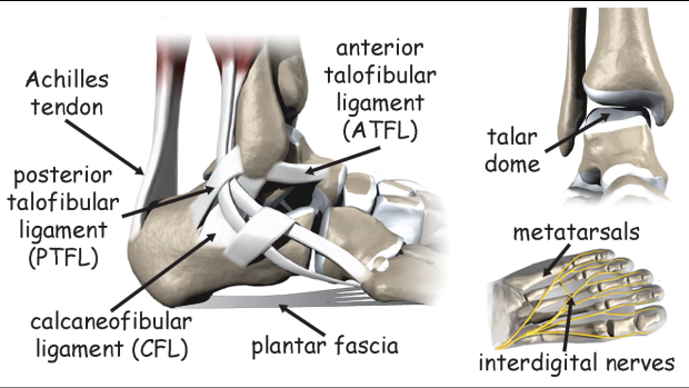
Anatomy of the ankle. Image credit
Lateral Ankle Sprain
Foot and Ankle Exam
One of the most common causes of foot pain is a lateral ankle sprain. In the mildest presentations, the only structures to be injured are the lateral ankle ligaments, usually in the number or one or two. However, if the damage is more severe, we can observe lesions of all the lateral ligaments plus other structures like the talar dome, the base of 5 th metatarsal and the tibio-fibular syndesmosis.
The approach is to check for the integrity of the lateral ligaments first and if all the three ligaments are injured, then we will check the other structures.
Inspection and Palpation
First, have the patient lying down supine with the knee bent on the affected side.
Then, observe the lateral aspect of the foot and ankle for hematomas or bruises.
Then, locate the three lateral ligaments and palpate along their course for crepitus and tenderness.